Translate this page into:
Osteocartilaginous choristoma of palatine tonsil: A rare entity
*Corresponding author: Dr. Mukta Pujani, MD Pathology, ESIC Medical College Faridabad, Haryana, India. drmuktapujani@gmail.com
-
Received: ,
Accepted: ,
How to cite this article: Batra A, Dhingra S, Pujani M, Khandelwal A, Singh K. Osteocartilaginous choristoma of palatine tonsil: A rare entity. Ann Natl Acad Med Sci (India) 2024;60:278-81. doi: 10.25259/ANAMS-2023-8-14-(1018)
Abstract
Choristoma is the presence of normal tissue in an abnormal anatomical location. The presence of mature cartilage in the tonsil represents a choristoma as it is not a normal constituent of the tonsil and is a very rare entity. More than 70% of lingual choristomas occur in females; the tongue is the most common site, accounting for 80% of the cases. Osseous choristoma of the tongue is a rather rare entity, with less than 100 cases reported in the literature. We hereby report an osteocartilaginous choristoma of the palatine tonsil in a 42-year-old male patient who presented with a sore throat and difficulty in swallowing, for which he underwent tonsillectomy. Choristoma was an incidental discovery. Histopathology features were consistent with chronic tonsillitis along with incidental occurrence of hyaline cartilage and bone. As choristoma is a benign tumor that usually does not require any further treatment after simple excision, therefore no further treatment was given. The patient is currently under a 2- year follow-up and does not show any signs of recurrence. This case creates awareness about this rare entity among pathologists and clinicians so that overzealous and unnecessary treatment is avoided.
Keywords
Choristoma
ectopic tissue
heterotopic tissue
Palatine tonsil
oral cavity
INTRODUCTION
The neck region is known for various embryological anomalies on account of its complex development.1 Choristoma is a developmental anomaly of the second pharyngeal arch,2 which refers to the presence of histologically normal tissue or cells at an abnormal location. Cartilaginous choristomas occur in young females and are presented as firm to hard, painless nodules reported in the head and neck region.1
Osteocartilaginous choristoma, consisting both osseous and cartilaginous tissues, is extremely rare in the oral cavity with only 10 cases documented in the English literature. In the oral cavity, the dorsum of the tongue is the usual site among women in their fifth decade of life. Etiology remains uncertain.3 Osteo-cartilaginous choristoma is very rare in tonsils.4–6
Osteocartilaginous choristoma is a well-defined swelling containing both osseous and cartilaginous tissues4. In the oral cavity, it is extremely rare with only nine cases reported in the English-language literature. At this site, these lesions especially involve the dorsum of the tongue in women from the 5th decade of life4. Etiology remains uncertain5 and histopathology is characterized by a mass of osseous and cartilaginous tissues4. Management is based on complete surgical resection.6 Osteocartilaginous choristoma is a well-defined swelling containing both osseous and cartilaginous tissues4. In the oral cavity, it is extremely rare with only nine cases reported in the English-language literature. At this site, these lesions especially involve the dorsum of the tongue in women from the 5th decade of life4. Etiology remains uncertain5 and histopathology is characterized by a mass of osseous and cartilaginous tissues4. Management is based on complete surgical resection6 Osteocartilaginous choristoma is a well-defined swelling containing both osseous and cartilaginous tissues4. In the oral cavity, it is extremely rare with only nine cases reported in the English-language literature. At this site, these lesions especially involve the dorsum of the tongue in women from the 5th decade of life4. Etiology remains uncertain5 and histopathology is characterized by a mass of osseous and cartilaginous tissues4. Management is based on complete surgical resection6 Osteocartilaginous choristoma is a well-defined swelling containing both osseous and cartilaginous tissues4. In the oral cavity, it is extremely rare with only nine cases reported in the English-language literature. At this site, these lesions especially involve the dorsum of the tongue in women from the 5th decade of life4. Etiology remains uncertain5 and histopathology is characterized by a mass of osseous and cartilaginous tissues4. Management is based on complete surgical resection6
CASE REPORT
A 42-year-old male presented to the clinic with recurrent episodes of sore throat for the past 2 years, along with difficulty in swallowing and snoring for the past 1 month. There was no history of ill-fitting dentures or any other dental problem. On examination, bilateral tonsils were enlarged with firm to hard areas on palpation. A provisional diagnosis of chronic tonsillitis with tonsilloliths was made. The dental examination was within normal limits. The rest of the head and neck regions did not reveal any abnormality. Bilateral tonsillectomy was performed, and the specimen was sent for histopathological examination. Written consent was obtained from the patient.
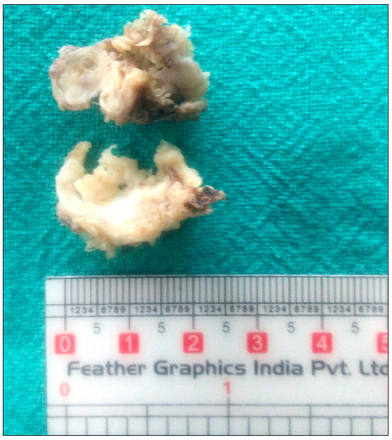
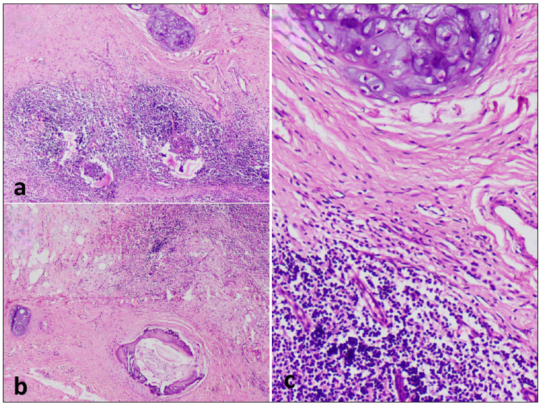
We received specimens of bilateral tonsils measuring 2 × 1 × 0.5 cm and 2 × 1.5 × 0.5 cm. Both cut surfaces were gray-white with few chalky white areas [Figure 1]. Histopathological examination revealed lymphoid hyperplasia, fibrosis areas, and mature hyaline cartilage islands [Figures 2a], occasional focus of mature bone formation [Figure 2b] and high power view of the cartilage [Figure 2c]. A diagnosis of osteo-cartilaginous choristoma of the tonsil was rendered. As choristoma is a benign tumor that usually does not require any further intervention or treatment after simple excision, and so no further treatment was given. The patient is currently under a 2-year follow-up, without any signs of recurrence.

- Gross photograph of cut surface of bilateral tonsils revealing gray white with few chalky white areas.

- Microphotograph revealing (a) lymphoid hyperplasia, areas of fibrosis, and islands of mature hyaline cartilage (H&E; 40x); (b) occasional focus of mature bone formation (H&E; 40x) and (c) high power view of cartilage (H&E; 100x). H&E: Hematoxylin and eosin stain.
DISCUSSION
Choristomas are benign lesions characterized by the presence of histologically normal tissue in abnormal locations due to developmental defects.1 The age of diagnosis ranges from 10 to 80 years.7 Cartilaginous choristomas of the oral cavity are rare, and most of the choristomas are osseous with a predilection for the tongue followed by buccal mucosa and soft palate.4,8 Chondroid choristomas of the tongue mostly occur in females, while no sex predilection has been observed in palantile tonsil.7 Osseous choristoma of the tongue is a rather rare entity, with less than 100 cases reported in the literature. So far, very few cases of cartilaginous choristomas of the tonsils have been reported.
Usually, they are observed as incidental findings during histopathological examination of tonsillectomies performed due to chronic tonsillitis. Erkilic et al. (2002) reported an incidence of 3% on tonsillectomy specimens.9 Sulhyan et al. (2016) in their study on tonsillar lesions found the incidence to be 2.84%.10 The present case is similar to the case by Pandey et al.2 (2012), where patient presented with recurrent episodes of chronic tonsillitis and was diagnosed as choristoma later.
Several hypotheses have been proposed to explain the pathogenesis of choristoma. Haemel et al. (2008) concluded that it arises from mesenchymal progenitor cells having multilineage potential, which were able to differentiate into various mesenchymal cell types.11 Lindholm et al. (1973) proposed that chemical or physical changes induced by chronic inflammatory processes could be responsible for the liberation of osteogenic substances, which stimulate heterotopic proliferation of cartilage.12 The lateral part of the second pharyngeal arch leads to the development of tonsils. Partihiban et al. (2011) postulated that choristomas of the tonsil arise from embryological anomaly of the second pharyngeal arch, which leads to the occurrence of abnormal mesenchymal tissue in the tonsil.13
Table 1 depicts the spectrum of choristoma cases of the oral cavity over the last decade.
| Authors | No. of cases | Age | Gender | Site | Type |
|---|---|---|---|---|---|
| Goncalo et al.14 (2024) | 1 | 72 | F | Tongue | Cartilaginous |
| Ali et al.15 (2024) | 1 | 30 | M | Nasopharynx | Cartilaginous |
| Shamloo et al.16 (2023) | 1 | 51 | F | Palate | Osseous |
| Pol et al.17 (2022) | 2 |
30 52 |
F M |
Soft palate Gingiva |
Osseous Osseous |
| Amaral et al.18 (2022) | 1 | 38 | Tongue | Osteocartilaginous | |
| Arimoto et al.19 (2021) | 1 | 11 | F | Tongue | Osseous |
| Gautam et al.20 (2021) | 1 | 38 | F | Tonsil | Cartilaginous |
| Bairwa et al.6 (2018) | 1 | 11 | M | Tonsil | Osteocartilaginous |
| Camara et al.3 (2017) | 1 | 59 | F | Tongue | Osteocartilaginous |
| Yoshimura et al.21 (2018) | 1 | 7 | M | tongue | Osseous |
| Qin et al.22 (2014) | 1 | 8 | M | Tongue | Osteocartilaginous |
| Meram et al.23 (2017) | 1 | 3 months | F | Skull | Osteocartilaginous |
Cartilage choristoma needs to be differentiated from metaplasia. Metaplasia is characterized histologically by diffuse calcific deposits and scattered chondrocytes in various stages of maturation single or as foci, whereas only mature tissue is present in choristoma.4
Excision remains the mainstay of treatment. In view of high recurrence rates in certain extraoral cases, excision should involve the removal of perichondrium as it has the potential to develop new cartilage, if left behind.7
CONCLUSION
Choristomas although rare entity, are usually discovered incidentally and are of academic interest only. They may be confused with true neoplasms if large in size or tonsilloliths in case of osseous or chondro-osseous choristoma. Moreover, the pathologist must be aware of this entity to avoid misdiagnosis of a benign incidental finding as some neoplasm.
Authors’ contributions
M.P, A.K: Idea and design; A.B, K.S: Data acquisition; S.D, A.K: Analysis; S.D, K.S: Interpretation of findings; A.B: Preparation of manuscript; M.P: Critical revision.
Ethical approval
Institutional Review Board approval is not required.
Declaration of patient consent
The authors certify that they have obtained all appropriate patient consent.
Financial support and sponsorship
Nil.
Conflicts of interest
There are no conflicts of interest.
Use of artificial intelligence (AI)–assisted technology for manuscript preparation
The authors confirm that there was no use of artificial intelligence (AI)-assisted technology for assisting in the writing or editing of the manuscript and no images were manipulated using AI.
References
- Cartilaginous choristoma of the tonsil: Three case reports. Iran J Otorhinolaryngol. 2015;27:325-8.
- [PubMed] [Google Scholar]
- Osteocartilaginous choristoma of the tongue: A case report and review of the literature. J Oral Diag. 2017;2:e20170009.
- [Google Scholar]
- Osteo-cartilagious choristoma of tonsil. J Evol Med Dent Sci. 2014;3:8916-7.
- [CrossRef] [Google Scholar]
- Osteocartilaginous choristoma of tonsil: A report of two cases. Arch Med Health Sci. 2019;7:78-80.
- [CrossRef] [Google Scholar]
- Osteocartilaginous choriostoma of palatine tonsil: A rare hidden entity. Iran J Pathol. 2018;13:471-3.
- [PubMed] [PubMed Central] [Google Scholar]
- Cartilaginous choristoma of tonsil: A hidden clinical entity. J Oral Maxillofac Pathol. 2013;17:292-3.
- [CrossRef] [PubMed] [Google Scholar]
- Histological features in routine tonsillectomy specimens: The presence and the proportion of mesenchymal tissues and seromucinous glands. J Laryngol Otol. 2002;116:911-3.
- [CrossRef] [PubMed] [Google Scholar]
- Histopathological spectrum of lesions of tonsil- A 2 year experience from tertiary care hospital of Maharashtra, India. Int J Med Res Rev. 2016;4:2164-9.
- [CrossRef] [Google Scholar]
- Heterotopic salivary gland tissue in the neck. J Am Acad Dermatol. 2008;58:251-6.
- [CrossRef] [PubMed] [Google Scholar]
- Histodynamics of experimental heterotopic osteogenesis by transitional epithelium. Acta Chir Scand. 1973;139:617-23.
- [PubMed] [Google Scholar]
- Choristoma of the palatine tonsil. A case report. Anatomica Karnataka. 2011;5:50-2.
- [Google Scholar]
- Oral A report of an unusual case of cartilaginous choristoma of the tongue and review. Surgery. 2024;17:147-51.
- [Google Scholar]
- Cartilaginous choristoma of the oral cavity: A rare presentation in the nasopharynx. Case Rep Med. 2024;2024:4506082.
- [Google Scholar]
- Osseous choristoma: Report of a case on the palate and a literature review. Clin Case Rep. 2023;11:e8355.
- [CrossRef] [PubMed] [PubMed Central] [Google Scholar]
- Osseous choristoma: Report of two cases in oral cavity. Indian J Pathol Oncol. 2022;9:279-81.
- [CrossRef] [Google Scholar]
- Choristoma of the dorsum of the tongue: A case report. International Archives of Otorhinolaryngology.. 2022;26:40.
- [Google Scholar]
- Tongue osseous choristoma in an 11-year-old female: A case report and literature review focusing on pediatric cases. Case Rep Dent. 2021;2021:8021362.
- [CrossRef] [PubMed] [PubMed Central] [Google Scholar]
- Cartilaginous choristoma of tonsil: A hidden clinical entity. Kathmandu Univ Med J (KUMJ). 2021;19:528-30.
- [CrossRef] [PubMed] [Google Scholar]
- Osseous choristoma of the tongue: A case report with dermoscopic study. Mol Clin Oncol. 2018;8:242-5.
- [CrossRef] [PubMed] [PubMed Central] [Google Scholar]
- [Tongue osteocartilaginous choristoma: A case report] Hua Xi Kou Qiang Yi Xue Za Zhi. 2014;32:96-8.
- [CrossRef] [PubMed] [PubMed Central] [Google Scholar]
- Benign malformation lesion of the skull: Hamartoma with ectopic elements or choristoma? Turk Patoloji Derg. 2017;33:262-7.
- [CrossRef] [PubMed] [Google Scholar]


