
Translate this page into:
Anthropometric Study of Proximal Femur Geometry and Its Clinical Application
COL. SANGHAM LAL MEMORIAL ORATION delivered during the NAMSCON 2018 held at the Mahatma Gandhi Medical College & Research Institute, Puducherry.
Correspondence: Dr. Ramchander Siwach, Senior Professor and Head, Department of Orthopaedics, Pt. B.D. Sharma PGIMS, Rohtak, Haryana, India. Email: rcsiwach.bps@gmail.com.
Abstract
The implants for fixation of proximal femur fractures and joint replacements have been designed taking into consideration of the anthropometry of the western population which vary from other ethnic groups. The present study aimed to study the morphology of the upper end of femur in relation to its various diameters and angles and compare the external and internal geometry of proximal femur as obtained from radiographs, with actual measurements on cadaveric specimens in Indian population. Seventy five pairs (150 bones) of cadaveric femora were studied morphologically and radiologically using standardized techniques to obtain various anthropometrics measurements. These values were compared with those reported in the literature for Hong Kong Chinese, Caucasian, Chinese and Western populations. Data were found to be quite different from them. It is proposed that implants designed for Western populations should be used judiciously and future implants be designed to match the morphology of the Indian bones.
Keywords
Anthropometry
proximal femur
cadaveric
Indian population.

Introduction
Operations on the proximal femur are one of the commonest in orthopaedic surgical practice. The aim of these operations is to remove pathology and restore anatomy to the normal, as far as possible. The implants for fixation of proximal femur fractures and joint replacements have been designed taking into consideration of the anthropometry of the western population which vary from other ethnic groups (1, 2). The standard commercially available marketed prostheses sometimes may not be the best fit to all subjects because of the large anatomic variation among different populations (2). The osteological parameters of the proximal femur are very important for the design of suitably sized prostheses of total hip replacement (THR), especially for cementless implantation (3). Orthopaedic surgeons always stress the need for a proper implant-patient match in hip joint replacements to avoid post-operative complication of mismatch which may affect the outcome of the operation (2).
Whereas what is normal has been standardized for Caucasians and Chinese (4-6), data for Indians are lacking. Since build, physique, habits and genetic make up vary markedly in different ethnic groups, it is possible that anthropometric dimensions described as normal for proximal end femur for Westerners might be quite different from those encountered amongst Indians. The present study was conducted with aim to remove the lacuna of information about proximal femoral geometry in Indian people and evaluate its impact on implant design. The present study aimed to investigate the morphology of the upper end of femur in relation to its various diameters and angles and compare the external and internal geometry of proximal femur as obtained from radiographs, with actual measurements on cadaveric specimens. The clinical application of the various geometric data, with the implants available, for osteosynthesis of the upper end of the femur and hip arthroplasty was also studied.
Materials and Methods
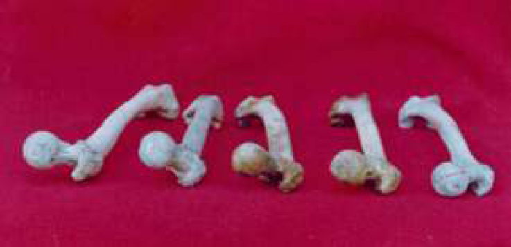
The study was conducted on 150 adult cadaveric femora (Fig. 1). Specimens that showed osseous pathology or previous fractures were excluded from the study. With the help of forensic expert these 150 adult cadaveric femora were differentiated into male and female femora, and their approximate age was determined. We did study on femora of adult group (age approximately between 20-80 years). Roentgenograms of 75 pairs of near identical specimen were taken in antero-posterior and lateral views using a precise standardized technique.
The specimens were placed directly over the cassette so the magnification would be insignificant. The distance between the X-ray source and the film was 1.2 m and the beam was centered on the lesser trochanter with the femur lying in neutral rotation. For lateral view without moving the femur, the X-ray source was rotated through 90° in the vertical plane, the distance between the source and the film remaining the same. Then the femur was kept on a sponge of the 2 feet length, 10" breadth and 8" height; the X-ray cassette was kept touching the femur, with one technician holding the cassette after wearing a lead apron. But on these lateral views the whole neck profile was not clear due to superimposition of greater trochanter. So to avoid this problem we kept the femur directly on the cassette in frog leg view position holding the condyles of the femur. In this view the neck profile of femur was clear.

- Cadaveric femora of 150 adults.
Morphological Study
The standard extracortical and endosteal dimensions were determined by direct measurement of cadaveric specimens. These measurements were done with the help of vernier caliper and goniometer (Fig. 2). With the help of vernier caliper we measured femoral head diameter, femoral head length, effective neck length, femoral neck diameter and canal width 20 mm above lesser trochanter, at level of lesser trochanter and 20 mm below lesser trochanter. With the help of goniometer neck shaft angle and angle of anteversion were measured.

- Showing various extra cortical dimensions in cadaveric femur and instruments used for various measurements.
Femoral head diameter: The distance between the two extreme points of head was measured.
-
Femoral head length: Radius of femoral head is not equal superiorly and inferiorly due to its placement (Fig. 3):
Maximum femoral head length - From the center of the femoral head to the periphery of femoral head along the articular cartilage border where it is maximum (superior);
Minimum femoral head length - From the center of femoral head to the periphery of femoral head along the articular cartilage border where it is minimum (inferior).
Effective neck length: (i) Maximum effective neck length - The length of neck where it is maximum was measured along the calcar (inferior); (ii) Minimum effective neck length - The length of neck where it is minimum was measured (superior).
Neck diameter: (i) Anteroposterior neck diameter - The distance between the two extreme points in middle of neck from the center point of intertrochanteric line to base of head in anteroposterior plane was measured; (ii) Superioinferior neck diameter - The distance between the two extreme points in middle of neck in superioinferior plane was measured (saggital plane).
Canal width, 20 mm above lesser trochanter. It was marked with a sketch pen 20 mm above and parallel to the horizontal axis passing through the center of lesser trochanter.
Canal width at level of lesser trochanter: A horizontal line was drawn through the center of lesser trochanter on anterior side.
Extracortical width 20 mm below lesser trochanter: It was marked with a sketch pen 20 mm below and parallel to the horizontal axis passing through the center of lesser trochanter.
Neck shaft angle: Center of head of femur was marked. Then mid point of the neck was marked by measuring the width of the narrowest portion of the neck and dividing by two. The line from center of the head of femur through the center of the neck was drawn. A line through the centre of the diaphysis of the femur was drawn. These two lines intersected each other. The angle between the two was measured.
Angle of anteversion: The center of the neck between its anterior and posterior surfaces was determined at two different points on the neck, as viewed from above. A line was drawn connecting these two points (Fig. 4). The femur was placed on a smooth level, horizontal surface so that it rested on three points, namely, the posterior aspect of the two femoral condyles and the posterior aspect of the greater trochanter. The goniometer was placed on the block of wood on which femur was rested. One arm of goniometer was opened and rotated till it was corresponding to the line connecting 2 center points marked on the neck of femur. The angle thus formed was read directly from goniometer, the eye kept on a level with the axis of the neck.

- Showing femoral head length in superior and inferior quadrant in cadaveric femur.
- Showing variations in angles of anteversion in cadaveric femur.
Radiological Study
A center point was marked at the level of isthmus. Second point was marked at the center of femur 3 cm above isthmus and third point was marked at the center of femur 3 cm below isthmus, a line connecting these 3 points was drawn and extended upwards and downwards.
With the help of scale we measured femoral head offset, femoral head diameter, femoral head position, neck diameter, canal width 20 mm above lesser trochanter, canal width at level of lesser trochanter, canal width 20 mm below lesser trochanter, endosteal width at the isthmus and extracortical width at the isthmus and isthmus position.
Femoral head offset: The distance between the center of head of femur and vertical axis drawn on femur.
Femoral head diameter: Two points were marked at the maximum distance on the head and the distance between the two was measured.
Femoral head position: It is the distance between the center of head and the horizontal line drawn through center of lesser trochanter.
Neck diameter: The width of the narrowest portion of the neck was measured.
Canal width, 20 mm above lesser trochanter: Two points were marked 20 mm above the lesser trochanter at maximum intracortical area and the distance between them was measured.
Canal width at level of lesser trochanter: Two points were marked at the level of lesser trochanter at maximum intracortical area and the distance between them was measured.
Canal width 20 mm below lesser trochanter: Two points were marked 20 mm below the lesser trochanter at maximum intracortical area and the distance between them was measured.
-
Endosteal width at the isthmus: The narrowest portion of the medullary canal is called isthmus. Two points were marked at this level at maximum intracortical area and the distance between them was measured.
Extracortical width at the isthmus: Two points were marked at the above level at the maximum extracortical area and the distance between them was measured.
Isthmus position: The distance between the isthmus and the center of lesser trochanter was measured.
Neck shaft angle: Center of head was marked. The mid point of the neck was located by measuring the width of the narrowest portion of the neck and dividing by two. The line connecting the center of head of femur through the center of the neck was drawn and extended to meet the vertical axis marked on femur. The angle formed between these two lines was measured by goniometer.
Canal flare index (CFI): It is defined as the ratio of the intracortical width of the femur at a point 20 mm proximal to the lesser trochanter to that at the medullary isthmus, allowing us to classify the femur into three general shapes: Normal, Stove pipe and Champagne flute.
Various anatomical representation and radiological measurement parameters are well depicted in Fig. 5.

- Anatomical representation and radiographic measurement on femoral radiographs.
Clinical Correlation
The implants used for osteosynthesis and arthroplasty were inserted in these bones, as described in their respective operative steps. The operations performed were dynamic hip screw, dynamic condylar screw, cancellous screws, and blade plate both 95° and 130°, for osteosynthesis, and femoral endoprosthesis for arthroplasty. In the cases of femoral arthroplasty the clina clay was used as cement to assess the cement mantle. After performing operations these bones were examined morphologically as well as radiologically as described above.
The comparison was done of both radiological and morphological measurements of lengths, diameters and angles. These parameters were correlated with the lengths, diameter and angles of standard implants available in the market for fixation of fracture trochanter, fracture neck femur and arthroplasty of hip. The standardization of the implants have been done taking into account that the present parameters of implants are acceptable in western bone mass, the percentage of volume occupied by these implants in western bone was compared, by the percentage of volume occupied by these implants in our femoral bone. And taking these as standard, modifications in implants size will be suggested in accordance to the anthropometric study of our race femora.
Results
Table 1 shows the average values of the morphological parameters studied, their standard deviation, minimum and maximum values and Table 2 shows the radiological aspect of all the morphological measurements. Table 3 shows comparison with Western and Asian (Chinese and Caucasians in Hong Kong). The volume of implants in the femoral head was calculated using d2 /4 x l where d is diameter of femoral head and l is length of implant [l =2/3 d- 10 mm (subchondral bone left)]. Table 4 depicts the percentage of femoral head volume occupied by various implants in different populations. Cross-sectional area of femoral neck is calculated by Formula p d2/4 (d = diameter). Table 5 represents the percentage of cross-sectional area of neck p d2 /4 occupied by various implants in different populations.
| Dimensions | No. | Average | Minimum (mm) | Maximum (mm) | Standard deviation (mm) |
|---|---|---|---|---|---|
| Femoral head diameter | 150 | 43.95 | 35.4 | 50.0 | 3.06 |
| Femoral head length | |||||
| Maximum (superiorly) | 150 | 36.9 | 24.4 | 49.2 | 4.11 |
| Minimum (inferiorly) | 25.5 | 16.0 | 36.5 | 4.26 | |
| Effective neck length | |||||
| Maximum (superiorly) | 150 | 37.23 | 26.5 | 50.5 | 4.65 |
| Minimum (inferiorly) | 22.69 | 16.3 | 39.2 | 3.65 | |
| Neck diameter | |||||
| Anteroposterior | 150 | 24.90 | 18.7 | 34.4 | 2.94 |
| Superoinferior | 31.87 | 23.3 | 40.9 | 2.91 | |
| Extracortical width, 20mm above lesser trochanter | 150 | 50.24 | 39.7 | 63 | 4.81 |
| Extracortical width at level of lesser trochanter | 150 | 40.44 | 29.8 | 52.6 | 4.67 |
| Extracortical width 20mm below lesser trochanter | 150 | 30.70 | 22.1 | 36.6 | 3.13 |
| Neck shaft angle (°) | 150 | 123.5° | 114° | 136° | 4.34 |
| Angle of anteversion (°) | 150 | 13.68° | 0° | 36° | 7.92 |
| Dimensions | No. | Average | Minimum (mm) | Maximum (mm) | Standard deviation (mm) |
|---|---|---|---|---|---|
| Femoral head offset | 75 | 38 | 29 | 47 | 5.52 |
| Femoral head diameter | 75 | 43.53 | 38 | 49 | 3.40 |
| Femoral head position | 75 | 50.15 | 41 | 62 | 4.80 |
| Neck diameter | 75 | 29.5 | 24 | 35 | 3.19 |
| Canal width, 20 mm above lesser trochanter | 75 | 43.5 | 33 | 53 | 4.37 |
| Canal width at level of lesser trochanter | 75 | 23.8 | 18 | 30 | 3.20 |
| Canal width 20 mm below lesser trochanter | 75 | 16.57 | 12 | 21 | 1.99 |
| Endosteal width at the isthmus | 75 | 10.11 | 6 | 15 | 1.90 |
| Extracortical width at the isthmus | 75 | 24.42 | 20 | 30 | 2.54 |
| Isthmus position | 75 | 112.92 | 87 | 128 | 10.58 |
| Neck shaft angle (°) | 75 | 123° | 118° | 140° | 4.29 |
| Average Dimensions | Present study (Indian) | Western (6,7) | Caucasian (5) | Hongkong (Chinese) (5) |
|---|---|---|---|---|
| Femoral head offset | 38 | 43 | - | - |
| Femoral head diameter | 43.53 | 46.1 | 46 | 45 |
| Femoral head volume (mm3) | 29618.55 | 34181.41 | 30744.48 | 26782.67 |
| Length of implant in femoral head (mm) | 19.3 | 20.73 | 19.67 | 18.33 |
| Femoral head position | 50.15 | 51.6 | - | - |
| Neck diameter | 29.5 | - | 33 | 31 |
| Canal width, 2 0mm above lesser trochanter | 43.5 | 45.4 | - | - |
| Canal width at level of lesser trochanter | 23.8 | 29.4 | - | - |
| Canal width 20mm below lesser trochanter | 16.57 | 20.9 | - | - |
| Endosteal width at the isthmus | 10.11 | 12.3 | - | - |
| Extracortical width at the isthmus | 24.42 | - | - | - |
| Isthmus position | 112.92 | 113. 4 | - | - |
| Neck shaft angle (°) | 123° | 124.7° | 136° | 135° |
| Angle of anteversion (°) | 13.68° | - | 7° | 14° |
| Cross-sectional area of femoral neck (mm2) | 633 | - | 778.92 | 660.12 |
| Different studies | 3 Cancellous screws | 3 Acinis screws | 2 Garden screws | DHS | Blade plates |
|---|---|---|---|---|---|
| Western | 6.03 | 6.99 | 6.88 | 7.44 | 4.39 |
| Caucasian | 6.37 | 7.38 | 7.26 | 7.85 | 4.64 |
| Asian (Hongkong Chinese) | 6.81 | 7.89 | 7.76 | 8.39 | 4.96 |
| Indian | 6.24 | 7.24 | 7.11 | 7.69 | 4.55 |
| Our study | 6.48 | 7.52 | 7.39 | 7.99 | 4.72 |
| Different studies | 3 Cancellous screws | 3 Acinis screws | 2 Garden screws | DHS | Blade plates |
|---|---|---|---|---|---|
| Caucasian | 12.77 | 14.81 | 14.56 | 15.75 | 9.31 |
| Asian (Hongkon g Chine se) | 15.07 | 17.48 | 17.18 | 18.58 | 10.98 |
| Our study | 15.72 | 18.23 | 17.92 | 19.38 | 11.45 |
Discussion
There are considerable variations in the femoral geometry of populations across different geographical locations and ethnic groups (3). Implants for fixation of proximal femur fractures have been designed taking into consideration of the anthropometry of the western population which varies from those of other ethnic groups (1). Similarly the standard commercially available marketed prostheses sometimes may not be the best fit to Indian patients because of the large anatomic variation (7). So the present study aimed to report proximal femoral geometry in Indian population and evaluate its impact on implant design.
In our ethnic race on radiological measurement average femoral head offset was 38 mm as compared to 43 mm in western literature. Similarly femoral head diameter in present study was 43.53 mm as compared to 46.1 mm in western literature. This shows that our skeleton is smaller than the western one. So in consideration of clinical importance of this parameter we shall have to think of smaller implants for osteosynthesis and may be a smaller size of endoprosthesis in few of our bones, especially in females. This dimension is also of clinical significance in acetabular cup size and the number of screws to be used in osteosynthesis of fracture neck of femur. In our set up smaller acetabular cup and lesser number of screws and smaller implants as DHS, DCS and blade plates need to be designed. As there is difference between the size and shape of the proximal femur of our race and western race with respect to canal width at different levels, hence the implants made according to western race do not fit accurately in our bones. There has to be a close match between the dimensions of the femur and the implant prosthesis. Similarly on radiological measurement the average intramedullary width of isthmus in our race was 10.11 mm (maximum being 15 mm and minimum being 6 mm) as compared to12.3 mm in western literature (maximum being 18.5 and minimum being 8 mm). There is marked difference between the two races in this parameter. This parameter is of immense importance in choosing the right size of the stem of endoprosthesis and the diameter of intramedullary nails because this parameter is much less in our race as compared to western race. On radiological measurement in our race average neck shaft angle was 123° (maximum being 140° and minimum being 118°). In western literature average neck shaft angle was 124.7°, maximum being 154.5° and minimum being 105.7°. In Caucasian male average is 136° with maximum being 161° and minimum 120°. In Caucasian female average is 133° with maximum being 145° and minimum 115°. This dimension is of significance in angled implants such DCS, DHS, blade plate. This angle being lesser in our race, we should prefer implants of lesser angle to avoid their superior cut through in the femoral head and neck. On radiological measurement femoral head position average was 50.15 mm in the present study as compared to 51.6 mm in western population. There is no significant difference between the two, probably because there is not much difference between neck shaft angle of our and western race.
The percentage of cross-sectional area of neck occupied by three cancellous screws of 6.5 mm in the neck is 12.77% in Caucasian as compared to 15.72% in our study. Therefore, the volume of bone mass replaced by metal is more in our patients as compared to counterpart in west. The percentage of femoral head volume occupied by three cancellous screws is 6.48% in our study as compared to 6.37% and 6.03%, in Caucasian and western, respectively. So, the chances of union reduce when 3 lag screws of 6.5 mm diameter each are inserted in the already compromised head and neck of the femur, especially in females. Therefore, it is advisable to put only two screws in place of three. In case we need to put 3 cancellous screws, one should be put as a cantilever along the superior border of the neck, which will hold only in the trochanter and the head. If we reduce the thread diameter of cancellous screws to 6.0 mm then, the percentage of femoral head volume occupied by three cancellous screws in our race becomes 5.52% which is nearly ideal for our race. The main hold of the screw threads is in the head of the femur. The head length is more in the superior part, the average is 36.9 mm as compared to the inferior part where the average is 25.5 mm. The cancellous screws are available in 16 mm and 32 mm thread length. Considering these parameters the screw thread, especially the 32 mm thread length, will not cross the fractured site in subcapital and transcervical fracture neck of the femur which is the prime requisite for union. Regarding the 16 mm thread length, it will cross the fracture site in subcapital and transcervical fracture neck femur in normal head which has reasonably adequate head length superiorly. In the central and superior area there is otherwise ample space available for a good hold. Therefore, in adults, subcapital and transcervical fractures, the 32 mm thread length, 6.5 mm or 7 mm cancellous screws should preferably not be used. The 16 mm thread length hold is good if they are passed through the center of the neck or in the superior quadrant. In the inferior area the head length is small and if the 5-10 mm subchondral area is left, as recommended the chances of the threaded portion crossing the fracture site is minimal even with 16 mm threaded screws. Therefore, accuracy is of prime importance regarding the length of the screws as the margin of error is less.
The percentage of femoral head volume occupied by DHS is 7.99%, 7.69%, 8.39%, 7.85% and 7.44% in present study, Indian counterpart, Asian, Caucasian and western studies, respectively. Similarly percentage of cross-sectional area of neck occupied by DHS in our study is 19.38%, Asians 18.58% and Caucasian 15.75%. The compression of bone in the head and replacement of bone mass by metal produces a tamponade effect in head which has a large bearing on nonunion and avascular necrosis; which are the key complications of fracture neck of femur. Secondly, there are two kinds of barrels in DHS, long and short ones with length of 38 mm and 25 mm respectively, with the outer diameter of 12.6 mm each. So while using this kind of barrel, one has to be considerate in accordance with the fracture line, otherwise the barrel will be longer. This will further occupy more space upto the longer portion along with the neck length and will not allow the controlled collapse. In our study the average neck length is 32 mm and only the short barrel should be used, because in using a long barrel there is always a danger of barrel crossing the fracture site, thereby preventing compression at the fracture site and controlled collapse thereafter. Thirdly the thread length of the DHS screw is 22 mm, with the outer diameter of 12.5 mm, and shaft diameter of 8 mm. To put the 12.5 mm screw we have to tap for 12.5 mm which takes out a lot of bone mass both from the neck and the head. The head length in our series varies from 25.5 mm to 36.9 mm and for proper purchase of DHS screw 5-10 mm subchondral bone is to be left, resulting thereby that screw thread which is 22 mm in DHS will not cross the fracture site in subcapital fractures. Lastly in our study, the neck shaft angle was found to be 123° (average radiologically), though ranging from 118° to 140°. DHS is available in angles starting from 135° to 150° at the difference of 5°. From the above information it appears that the 135° angle is more and hence chances of the superior cut through of the implant are more. Otherwise one has to make the entry point at such a level on the lateral side of trochanteric area so that the tip of screw lies in the center or posteroinferior quadrant of the head. To achieve this valgus osteotomy will be needed simultaneously for a better approximation of femoral shaft with the plate, and thereby achieving an undesirable overall coxa valga in comparison to contralateral hip which may result in limb length discrepancy and avascular necrosis of head of femur. We conclude that for the Indian patients, the implant size should be reduced to 11.5 mm in place of 12.5 mm, then the cross-sectional area occupied in the neck will be 16.40% and the percentage of femoral head volume occupied will reduce to 6.76%, which are within the desirable limits. This should be done even with compromising the strength of the implant, to have better biology, which is the key factor for union and vascularity. The threaded portion should also be reduced from 22 mm to 15 mm, and the implant should also be available in 120°, 125° and 130° in accordance with our patients requirement. Also we should always procure the X-rays of both the hips to achieve same neck shaft angle peroperatively as it varies from person to person. Regarding the diameter and thread length parameters, same is true with DCS screw but its angle is acceptable.
In the present study, the CFI varies from 2.75 to 8.5. Depending on the CFI, the shape of the medullary canal, the normal canal type is 50%, champagne-fluted canal type in 38.46% and stovepipe canal type in 7.69%, and in a few cases do not fit in any of the types described above. The implants available are in variable lengths, ranging from 126 mm to 174 mm, with distal width ranging from 7 mm to 11 mm and anteroposterior thickness ranging from 6 mm to 8.5 mm. Our isthmus position ranges from 87 mm to 128 mm (average 112.92 mm) and the endosteal diameter at the level of isthmus varies from 6 mm to 15 mm (average 10.11 mm). Therefore, in the Indian patients only smaller prostheses are used, and in case of thin and lean patients (especially of younger age group) even CDH implants are good enough. At a distance of 20 mm above the lesser trochanter, the anteroposterior canal width was found to differ by 45.4%, when compared with a French population which can affect the mechanical stability of femoral stem (8). We recommend endoprosthesis having length ranging from 100 mm to 150 mm, distal width from 6 mm to 11mm and anteroposterior thickness from 5.5 mm to 8 mm. The incidence of intraoperative complications like splintering and fractures ranges from 4% to 21% (9-11). These are due to over-sized implants available that have been manufactured basically with western parameters. Most femoral stems are designed to extend to the isthmus of medullary canal, so that the component is stable and there is a 2 mm cement mantle around it. Therefore, only smaller sized implant, both in length as well as in thickness, with straight and polished stem, are preferred. To achieve these conditions the manufacturers should reduce the geometric measurements of endoprosthesis but at the same time should not compromise on strength of implant. Another important point in total hip arthroplasty is restoration of original position of the center of head along with the limb length equality and the restoration of the original balance of abductors (6). For this purpose femoral components are available in a long range of neck lengths for each separate stem size. To have a good muscle balance and limb length, the head offset is an essential component. There are various implants available with variable offsets ranging from 32.8 mm to 50 mm. In our study the head offset ranges from 29 mm to 47 mm (average 38 mm), hence in our patients 37.5 mm to 44 mm head offsets are suitable. So the clinical result of the above observations made in relation to the medullary canal geometry is, that the implant needs to be designed on the basis of anthropometric data available, along with other factors like age, sex and the environment, etc. This will minimise the preoperative and postoperative complications involved in total hip arthroplasty, although for a perfect match each implant needs to be customised.
Some other authors have also reported the assessment of geometry of proximal femur in Indian population and have suggested modifications in implants. Pathrot et al (1) advocated certain modifications in the presently available short cephalomedullary nail designs for them to better fit the anatomy of our subset of population: (a) two nails of 125° and 135°; (b) the medio-lateral angle at the level of 65 mm from the tip of the nail; (c) two femoral neck screw placements (35 and 45 mm from the tip of the nail); and (d) five different sizes of distal width for better fit in canal (9-13 mm). Maji et al also found variations in the morphology of the proximal femur between the Indian population and that of other countries, and advocated the need for standardizing THR implant sizes for the Indian population, especially for cementless implantation (3). Rawal et al observed a difference of 16.8% in the femoral head offset between Indian and Swiss populations, which can affect soft tissue tension and range of motion (7). Maheshwari et al also reported that when compared with the Western data, the femoral neck anteversion values were 3-12 degrees lower and the combined anteversion values were 3-5 degrees lower in Indian adults (11). The acetabulum anteversion values were comparable, but were skewed towards the higher side (11). But Saikia et al observed that the neck shaft angle and the femoral neck anteversion in individuals of North eastern region of India was 5-6 degrees more than the western literature (12).
Anthropometric studies for proximal femur in ethnic groups other than western population have reported significant differences. Pi et al reported that Chinese proximal femoral parameters are significantly different from Westerners (13). Compared with Westeners, the offset was smaller, while the neck shaft angle was significantly larger in Chinese population (13). Most parameters of the proximal femoral medullary cavity diameter were significantly smaller in Chinese population than those in Westerners (13). Atilla et al reported osteometry of the femora in Turkish individuals (14). They observed diverse features of femoral geometry in Turkish individuals compared to Western populations and advocated that these differences should be taken into account in the design and development of hip prostheses (14).
The observations in the present study have profound implications. Not only are western implants large in size, their angles, and orientations and thread length also mismatch Indian femora. Implants designed for western skeletons occupy much more space in the Indian femoral head and neck. A certain subset of Indian femora does not have any implant available to them as they are too small. Furthermore, a shorter neck length implies that the threads of cancellous or Garden screws used to fix neck fractures may not cross the fracture site thereby failing to provide compression and thus defeating the whole purpose of the surgery. If too much bone is replaced by metal a tamponade effect can ensue that may cause avascularity of femoral head, consequently resulting in nonunion of neck fractures and/or AVN. Since our heads are smaller, the threads of screws often fail to cross the fracture of neck of femur especially if the fracture is sub capital and the screw placement in the inferior quadrant of head. This means we must have screws with shorter thread lengths. In thin built and short individuals the neck may not have space enough to occupy the three 6.5 mm screws recommended for fixation of neck fractures. A smaller neck shaft angle implies that a DHS inserted through the classical entry portal using angled guide will either go into the superior quadrant or pull the fracture in valgus both of which are undesirable. We probably require DHS with smaller angles. Our recommendations for different implants are shown in Table 6.
| Implants | Presently available dimensions | Our recommendations |
|---|---|---|
| Cancellous screw | Thread diameter 6.5 mm | Thread diameter 6 mm |
| Thread length 16 mm, 32 mm | Thread length 16 mm | |
| Acinis screw (cannulated) | Thread diameter 7 mm | Thread diameter 6.5 mm |
| Thread length 16 mm, 32 mm | Thread length 16 mm | |
| Garden screw | Thread diameter 8.5 mm | Thread diameter 8 mm |
| Thread diameter 12.5 mm | Thread diameter 11.5 mm | |
| DHS screw | Thread length 22 mm | Thread length 15 mm |
| Barrel short and long | Barrel short | |
| Angles 135° to 150° (at difference of 5°) | Angles 120° to 150° (at difference of 5°) | |
| Blade plate | ‘U’ profile 6.5x16 mm | ‘U’ profile 6.5x12.5 mm |
| Angles 95° and 130° | Angles 95°, 120°, 125° and 130° | |
| Standard Gamma nail | Modified by Leung et al1 | |
| Mediolateral angle-10° | Mediolateral angle 4° | |
| Gamma nail | Length 200 mm | Length 180 mm |
| Diameter of distal shaft 12 mm, 14 mm and 16 mm | Diameter of distal shaft 11 mm and 12 mm | |
| and 12 mm | This modified nail is also suitable to our race femora | |
| Length 126 mm to 174 mm | Length 100 mm to 150 mm | |
| Distal width 7 mm to 11 mm | Distal width 6 mm to 11 mm | |
| Anteroposterior thickness 6 mm to 8.5 mm | Anteroposterior thickness 5.5 mm to 8 mm | |
| Modularity up to 10 mm | Modularity up to 15 mm | |
| Endoprosthesis | Head offset 32.8 mm to 50 mm | Head offset 37.5 mm to 44 mm |
| Stem type – -Banana shape -Straight -Rough and grooved -Flat back -Rounded -Rectangular cross section -Trapezoidal and diamond shaped |
Stem type – Polished, straight and trappered collarless stem is preferred. |
The implications of the study on arthroplasty operations can not be overemphasized as these are designed to reproduce the normal anatomy as far as possible. Orthopaedic surgeons always stress the need for a proper implant-patient match in hip joint replacements, in particular, for a cementless femoral stem (7). The complications of mismatch are aseptic loosening, improper load distribution, and discomfort (7). The clinical symptoms are due to the bone implant mismatch, which result in micromotion. There are studies, which highlight that these micromotions should be reduced to 14 micra or less, to prevent osteolysis and aseptic loosening (5). We agree with Roy et al that improved knowledge of the morphology of the proximal femora will assist the surgeon in restoring the geometry of the proximal femur during total hip arthroplasty and the data could be used as a guideline to design a more suitable implant for Eastern Indian population (2).
References
- Assessment of the geometry of proximal femur for short cephallomedullary nail placement : an observational study in dry femora and living subjects. Indian J Orthop. 2016;50(3):269-276.
- [CrossRef] [PubMed] [Google Scholar]
- Evaluation of proximal femoral geometry in plain anterior- posterior radiograph in Eastern-Indian population. J Clin Diagn Res. 2014;8(9):AC01-AC03.
- [CrossRef] [PubMed] [Google Scholar]
- Investigating the morphology of the proximal femur of the Indian population towards designing more suitable THR implants. J Long Term Eff Med Implants. 2012;22(1):49-64.
- [CrossRef] [PubMed] [Google Scholar]
- Anatomy of the femoral neck and head, with comparative data from Caucasians and HongKong. Chinese Clin Orthop. 1980;152:10-16.
- [CrossRef] [Google Scholar]
- Do we need a special design of femoral component of total hip prosthesis in our patients? Indian J Orthop. 1999;33(4):282-284.
- [Google Scholar]
- The anatomical basis of femoral component design. Clin Orthop Rel Res. 1988;235:148-164.
- [CrossRef] [Google Scholar]
- Anthropometric measurements to design best-fit femoral stem for the Indian population. Indian J Orthop. 2012;46(1):46-53.
- [CrossRef] [PubMed] [Google Scholar]
- Cementless isoelastic total hip prosthesis: preliminary report on the first 21 5 consecutive cases. In: Morscher E, ed. In: The Cementless Fixation of Hip Endoprosthesis. New York: Springer-Verlag; 1984. p. :243.
- [CrossRef] [Google Scholar]
- Casual histogenesis and biomechanical discoveries as a basis for cementless fixation of hip endoprosthesis. In: Morscher E, ed. In: The Cementless Fixation of Hip Endoprosthesis. New York: Springer-Verlag; 1984.
- [CrossRef] [Google Scholar]
- A cementless titanium hip endoprosthesis system based on press-fit fixation: basic research and clinical results. Instr Course Lect. 1986;35:203-225.
- [Google Scholar]
- Femoral neck anteversion, acetabular anteversion and combined anteversion in the normal Indian adult population: a computed tomographic study. Indian J Orthop. 2010;44(3):277-282.
- [CrossRef] [PubMed] [Google Scholar]
- Anthropometric study of the hip joint in northeastern region population with computed tomography scan. Indian J Orthop. 2008;42(3):260-266.
- [CrossRef] [PubMed] [Google Scholar]
- Measurement of proximal femoral morphology and analysis of 500 cases in Hunan Province. Zhong Nan Da Xue Xue Bao YiXue Ban. 2013;38(9):925-930.
- [Google Scholar]
- Osteometry of the femora in Turkish individuals: a morphometric study in 114 cadaveric femora as an anatomic basis of femoral component design. Acta Orthop Traumatol Turc. 2007;41(1):64-68.
- [Google Scholar]


